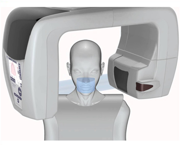
Cone Beam CT 3D Imaging
Cone Beam CT - 3D Imaging
WHAT IS CONE BEAM COMPUTED TOMOGRAPHY (CBCT)?
Your dentist can get a more complete image of your smile by utilizing cone beam 3D imaging. Dr. Erica Elannan offers this tool at Smile Nuvo in order to provide you with the highest level of dental care. For many years, dental professionals have relied on intraoral radiography, panoramic radiography and conventional tomography for diagnosis and treatment planning. These commonly used imaging modalities produce only two-dimensional and/or distorted images with superimposition of structures outside the area of interest. There has always been a need for three-dimensional (3-D) imaging but advanced technology has only recently become available. Cone Beam Computed Tomography (CBCT) uses a cone-shaped beam and digital processing to reconstruct a virtually distortion-free 3-D image using a single rotation in a sitting/standing position, similar to that of a panoramic radiograph. CBCT scanners are compact in size, capable of producing high-resolution 3-D imaging of hard tissues and are compatible with computer-aided imaging software for improved diagnosis and treatment planning.

WE ARE PROUD TO UTILIZE CONE BEAM 3D IMAGING TECHNOLOGY AT OUR PRACTICE
Cone beam 3D technology is an imaging system that provides your dentist and team with a three-dimensional image reconstruction of your teeth, mouth, jaw, neck, ears, nose and throat. We use dental cone beam 3D imaging to:
- Plan dental implant placement
- Evaluate jaws and face
- View the head and neck as a comprehensive whole Diagnose tooth decay (cavities) and other dental problems Detect endodontic problems and plan root canal therapy Analyze dental and facial trauma
- Plan and evaluate the progress of orthodontic treatment Visualize abnormal teeth
- Assess a TMJ disorder
- Benefits of CBCT imaging
- Imaging exposes patients to less radiation than traditional CT scans.
- The scan is fast and comfortable.
- A reduction in metal artifacts.
- A single scan produces a volume of images that can be viewed and manipulated. Clinicians can illustrate recommended treatment plans to patients using 3-D software. No superimposition and minimal distortion.
- Allows clinician to visualize internal anatomy that cannot be diagnosed externally. Lower cost for patient when compared to traditional CT.
- Enhanced communication with patients and colleagues.
To learn more about cone beam 3D imaging and how it helps us provide you with exceptional care, we invite you to contact our office at 602-834-8834 or inquire at your next appointment.
- Imaging exposes patients to less radiation than traditional CT scans.
- The scan is fast and comfortable to the patient.
- Reduction in metal artifacts.
- One scan produces a volume of images that can be viewed and manipulated.
- Clinicians can illustrate recommended treatment plans to patients using 3-D software.
- No superimposition and minimal distortion.
- Allows clinician to visualize internal anatomy that cannot be diagnosed externally.
- Lower cost for patient when compared to traditional CT.
- Enhanced communication with patients and colleagues.
To learn more about cone beam 3D imaging and how it helps us provide you with exceptional care, we invite you to contact our office at 602-834-8834 or ask us about it at your next appointment.
